Retinal Vein Occlusions (RVOs) refer to blockages in one or more of the veins that supply and return blood from the retina (the back of the eye). RVOs can lead to significant vision loss if not managed promptly. These are two main types of RVOs:
Central Retinal Vein Occlusion (CRVO): This occurs when the main vein (central retinal vein) that drains blood from the entire retina becomes blocked.
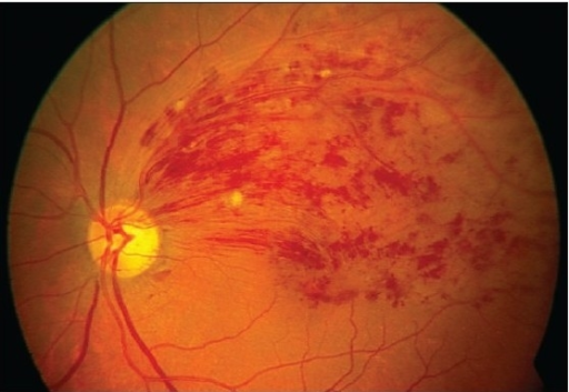
Branch Retinal Vein Occlusion (BRVO): This happens when one of the branch veins that drain a smaller area of the retina becomes blocked.
Causes:
- Arteriosclerosis: Hardening of the arteries at the back of the eye which in turn can push on the vein, causing it to become blocked.
- Thrombosis: This occurs when a blood clot forms within the vein, leading to a blockage.
- Compression: Pressure on the vein from bodily structures that lay next to the vein. Any abnormality in these structures can then push, or compress, the vein and thus leading to a blockage at the site of compression.
Inflammation: Conditions like vasculitis (inflammation of blood vessels) may cause a blocked retinal vein in the eye.
Risk Factors:
- Advanced age: RVOs are more commonly occurring in older adults, and this is believed to be likely due to the increased rate of risk factors including high blood pressure (hypertension) and hardening of arteries (arteriosclerosis) with age.
- Hypertension: Raised blood pressure levels can damage the blood vessels in the retina (back of the eye), leading to a retinal vein occlusion.
- Diabetes: Diabetes, raised blood sugar levels, can also cause damage to the blood vessels in the retina and thus, increases the risk of RVOs.
- Glaucoma: Raised eye pressures associated with glaucoma and compress, or push on, the retinal veins, therefore increasing risk of RVOs.
- Hyperlipidemia (high cholesterol): Raised cholesterol levels can contribute to the hardening of arteries (arteriosclerosis) and therefore increase the risk of RVOs.
Smoking: Tobacco use increases the risk of developing cardiovascular diseases (such as high blood pressure and high cholesterol) which in turn increases the risk of complications in the eye including retinal vein occlusions.
Symptoms:
Retinal vein occlusion (RVO) can cause various symptoms depending on the type (central or branch) and severity of the occlusion. Here are the common symptoms associated with retinal vein occlusion:
- Sudden Blurred Vision: Individuals may experience sudden onset of blurry or distorted vision in one eye. Vision may become significantly reduced, especially if the macula (central part of the retina responsible for sharp, central vision) is affected. The blurred vision may occur within a few hours or days after the blockage has occurred. You may notice that a particular portion of your vision has become suddenly worse (such as top or bottom half of your vision) – this can often happen when you have a superior (top) or inferior (bottom) branch of the retinal veins that have become blocked.
- Visual Field Defects: Some people may notice a sudden loss of peripheral vision or the appearance of a curtain or shadow in their visual field.
Diagnosis:
- Fundoscopy: This is done by your eye specialist where they will examine the back of the eye using a special magnifying lens. This is typically done to assess whether there is obvious bleeding (haemorrhages) or deposits (hard exudates) at the retina.
- Fluorescein Angiography: This involves injecting a special yellow dye into the bloodstream (usually via insertion of a cannula into the arm). The dye helps to highlight the blood vessels at the back of the eye and determine whether there are any leakages in the blood vessels.
- Optical Coherence Tomography (OCT): OCT scanning determines the retinal thickness. This imaging technique is particularly useful in determining baseline swelling at the back of the eye that often occurs after a retinal vein occlusion. Your eye specialist will also use OCT scanning at each visit to determine whether treatment is helping to reduce the retinal swelling. They can also compare baseline scans with your most recent scans.
Treatment:
- Management of Underlying Conditions:
- Control of Systemic Factors: This is particularly important to manage the underlying and often biggest contributing factor to RVOs. Targeted management of conditions such as high blood pressure, diabetes, and high cholesterol are crucial as they all can contribute to the risk and progression of RVOs. This can often be done in conjunction with your general practitioner (GP) where close monitoring may be required to adjust medication as necessary for the conditions mentioned above.
- Intravitreal Injections:
- Anti-VEGF Therapy: Medications such as ranibizumab (Lucentis), aflibercept (Eylea) are injected into the ‘jelly-like substance’ (vitreous cavity) of the eye. This is done by your friendly eye specialist and the medication works to reduce vascular endothelial growth factor (VEGF) which has been known to be responsible for the abnormal blood vessel growth and leakage that may occur after a vein blockage, or vein occlusion. The aim of anti-VEGF injections is to reduce the macular oedema (or retinal swelling) and thereby hoping to improve vision in the affected eye. In some cases, the main aim is to stabilise the vision and prevent it from worsening.
- Steroids: Eye injections of steroids like dexamethasone (Ozurdex) can be used to reduce inflammation and swelling in the retina. These are slow-release ‘implants’ that are injected into the ‘jelly-like’ substance in a very similar way to the conventional anti-VEGF injections however they generally last up to three months at a time. Patient eligibility is determined by the eye specialist during consultation.
- Laser Treatment:
- Laser Photocoagulation: This is used less frequently than anti-VEGF injections however your eye specialist may choose to perform this laser procedure alongside your anti-VEGF injections if you have complications including growth of new abnormal blood vessels at the site of the original vein occlusion.
- Surgical Interventions:
- Vitrectomy: In severe cases of RVOs that are complicated by bleeding in the ‘jelly-like’ substance (known as a vitreous haemorrhage), your eye specialist may decide that it would be best to perform a vitrectomy where the ‘jelly-like’ substance (vitreous) is removed and replaced with a saline solution, silicone oil, or gas bubble to maintain the eye’s shape. This surgery is done to remove the blood and scar tissue in the eye that may be causing reduced vision.
- Monitoring and Follow-Up:
Prognosis:
The prognosis for RVOs varies depending on the severity of the occlusion, presence of macular oedema, and timely commencement of treatment. Early diagnosis and intervention have been known to greatly improve visual outcomes and preventing irreversible vision loss. However, some individuals may still experience persistent visual impairment despite early treatment, particularly if there are further complications such as abnormal blood vessel growth or vitreous haemorrhages.
Patients with RVOs should work closely with their eye specialists to develop a tailored treatment plan that caters to their specific needs and general health medical history. Regular follow-ups are crucial to monitor the eye’s response to treatment and ensure the best possible visual outcome is achieved.